Can Soft Tumours Be Seen on X-Rays?
Soft tissue tumours are growths that develop in the body’s soft tissues, including muscle, fat, blood vessels, and connective tissue. While most soft tissue tumours are benign or non-cancerous, some can be malignant or cancerous. In addition, soft tumours can be difficult to see on X-rays. This is because they often don’t have the same density as bones or other hard tissues.
However, with the help of a trained radiologist, it is sometimes possible to see these types of tumours on an X-ray. This blog post will discuss the various methods used to detect soft tumours. We will also talk about the signs and symptoms of this condition. Finally, we will provide some tips on how to get the best results from your X-ray.
Symptoms
There are many different types of soft tissue tumours, which can occur anywhere in the body. These tumours can be benign or malignant, and they can vary significantly in size and appearance. Some common symptoms of soft tissue tumours include swelling, pain, and difficulty moving the affected area.
Treatment
Treatment for soft tissue tumours depends on the specific type of tumour, as well as its location and severity. Benign tumours can often be treated with surgery, while malignant tumours may require a combination of surgery, radiation therapy, and chemotherapy. The prognosis for patients with soft tissue tumours varies depending on the individual case. However, early diagnosis and treatment are often essential for the best possible outcome.
How do you detect Soft tumours?
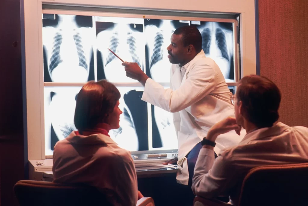
Now that we have discussed the basics of soft tumours let’s talk about how they are detected on X-rays. As we mentioned earlier, these tumours often don’t have the same density as other tissues. This means that they can be challenging to see on traditional X-rays. This depends on the site and the size of the tumour.
Specific locations, like in the lung, may be visible if the tumour is of adequate size (around 3 cm). However, small tumours or tumours located in the abdomen may not be visible on an x-ray. In such cases, if there is suspicion of a tumour, more detailed scans like CT, MRI or PET are performed. This is because these have a higher sensitivity to the detection of tumours.
However, there are a few different methods that can be used to detect soft tumours. One of these methods is called contrast-enhanced imaging. This method uses special dyes that help to make the tumour more visible on the X-ray.
Another method that can detect soft tumours is called computed tomography (CT). This type of scan uses a series of x-rays to create a three-dimensional image of the body. This image can then be viewed from different angles, which helps to identify the tumour.
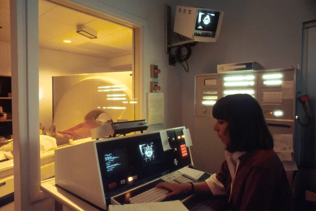
MRIs, or magnetic resonance imaging scans, are a type of imaging that uses powerful magnets and radio waves to produce detailed images of the inside of the body. MRIs are painless and don’t involve ionising radiation, making them a safe option for people of all ages.
MRIs can be used to detect a variety of conditions, including tumours. This is because it can provide detailed images of the soft tissues in the body, making it easier for doctors to locate tumours. In addition, MRIs can also help determine the size and shape of a tumour. In turn, this information helps to plan the best course of treatment.
A PET scan is an imaging test that uses radioactive tracers to produce detailed images of the body. PET scans are often used to detect cancer and evaluate the effectiveness of cancer treatment. The tracers used in PET scans are injected into the body and then absorbed by cells.
The amount of tracer uptake is then used to create body images. Cancerous cells tend to absorb more tracer than healthy cells, showing up as bright spots on the image. As a result, PET scans are beneficial for detecting small, early-stage cancers. They can also help determine if cancer has spread to other body parts.
With early detection, there is a better chance of preventing these tumours from becoming cancerous. If you are concerned that you or someone you know may have a soft tumour, it is essential to talk to your doctor. They will guide you with appropriate tests and give you the best course of treatment.
Thanks for reading😊


